Cellular Senescence Detection Comparison
Cellular senescence is considered crucial to many different research areas, especially since the recent discovery of the senescence-associated secretory phenotype (SASP). SASP is a known risk factor for malignant transformation and exploration into stem cell research has found a link between SASP and the aging phenomenon. The features of cellular senescence were summarized based on modern publications that have a high number of citations.
Various Senescent Cell from Various Indicators
| Product | Cellular Senescence Detection Kit - SPiDER-βGal | Cellular Senescence Plate Assay Kit - SPiDER-βGal | DNA Damage Detection Kit – γH2AX *Please Contact Us |
Nucleolus Bright Green / Red |
|---|---|---|---|---|
| Detection | Fluorescence | Fluorescence | Fluorescence | Fluorescence |
|
Wavelength (Ex/Em) |
500 - 540 nm / 530 - 570 nm | 535 nm / 580 nm | Green: 494 nm / 518 nm Red: 550 nm / 566 nm Deep Red: 646 nm / 668 nm |
Green: 513 nm / 538 nm Red: 537 nm / 605 nm |
| Indicator | SA-β-gal activity | SA-β-gal activity | Changes in DNA Damage | Changes in the Nucleolus |
| Detection method | Imaging Substrate: SPiDER-βGal |
Plate Assay Substrate: SPiDER-βGal |
Imaging Detection of γH2AX secondary antibody method |
Imaging Detection of the Nucleolus by RNA staining reagent |
| Instrument | Fluorescence Microscopy Flow cytometry |
Plate Reader | Fluorescence Microscopy | Fluorescence Microscopy |
| Sample | Live Cells, Fixed Cells | Live cells(Lysis of live cells) | Fixed cells | Fixed cells |
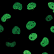
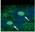
| Data |  |
 |
 |
 |
| Item# | SG03 | SG05 | Green: G265 / Red:G266 Deep Red: G267 |
Green: N511 / Red: N512 |
Feature of Cellular Senescence
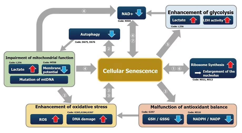
Evaluate senescent cells from various makers
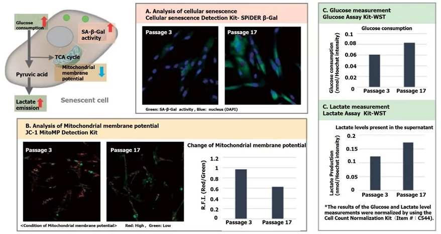
In WI-38 cells at different passages SA-ß-Gal activity, mitochondrial membrane potential, and cellular metabolism (Glucose and Lactate) were evaluated using each specific kit. In senescent cells, SA-ß-Gal activity was enhanced and mitochondrial membrane potential was reduced. Consumption of Glucose and lactate level in the supernatant measured as an indicator of metabolism was increased.